Datei:Determinants of Gastric Acid Secretion.svg

Größe der PNG-Vorschau dieser SVG-Datei: 800 × 550 Pixel. Weitere Auflösungen: 320 × 220 Pixel | 640 × 440 Pixel | 1.024 × 704 Pixel | 1.280 × 880 Pixel | 2.560 × 1.760 Pixel | 1.206 × 829 Pixel.
Originaldatei (SVG-Datei, Basisgröße: 1.206 × 829 Pixel, Dateigröße: 741 KB)
Beschreibung
| Beschreibung |
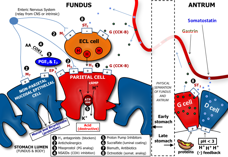
English: There are several neuronal, endocrine, and paracrine determinants of gastric acid secretion. Parietal cells in the fundus and body of the stomach are responsible for secretion of acid into the stomach. Major receptors on parietal cells that trigger acid release when activated include the M3 muscarinic, the H2 histaminic, and the CCK-B or gastrin receptor. The respective ligands for these receptors are acetylcholine, histamine, and gastrin. Acetylcholine is released from enteric neurons, histamine is released from enterochromaffin-like cells, and gastrin is released from G cells which are generally found in the antrum, or lower portion, of the stomach. Enterochromaffin-like (ECL) cells are generally found below the epithelial cell layer of the stomach lumen. ECL cells express M3 muscarinic, ST2 somatostatin, and CCK-B gastrin receptors. While M3 and CCK-B receptors trigger histamine release when activated, the ST2 somatostatin receptor inhibits histamine release. Thus, gastrin is pro-acid, and somatostatin is anti-acid secretion. Both somatostatin and gastrin are released primarily from cells in the antrum. G cells secrete gastrin, and D cells secrete somatostatin. D cells have a regulatory role over G cells in that the somatostatin that they release has an inhibitory influence on gastrin secretion from G cells. This serves to reduce futile simultaneous pro-acid and anti-acid signals to the fundus and body of the stomach. A major trigger for gastrin release is the presence of protein in the antrum: this serves to enhance acid secretion, which is necessary for the activity of pepsin, a major digestive enzyme in the stomach which is important for protein breakdown. In addition, D cells are also subject to regulation by gastric luminal contents. In particular, D cells release more somatostatin when the antral pH is low (i.e., when a highly acidic condition exists). This serves as a negative feedback when acid levels are high, as somatostatin will be secreted, enter the gastric bloodstream, and signal to ECL cells to reduce histamine release. Reduced histamine release will, in turn, reduce acid secretion from parietal cells. Finally, our gastric cells would be susceptible to damage from acid and pepsin if they had no protection. Protection of the gastric mucosal cells is in two forms, a physical barrier of viscous mucus, and a chemical defense of bicarbonate, which neutralizes acid. Protective prostaglandins, i.e., PGE2 and PGI2 activate EP3 receptors on non-parietal mucosal cells to enhance mucus and bicarbonate secretion. At the same time, EP3 receptors are activated on parietal cells to inhibit acid secretion. This is primarily a result of EP3's coupling to the G-protein, Gi, which reduces cyclic adenosine monophosphate (cAMP) levels. The proton pump in parietal cells is an exchanger of potassium and protons. Many portions of the physiology of gastric acid secretion are influenced by drugs, especially those used to treat peptic ulcer disease (PUD) and gastroesophageal reflux disease (GERD). The drug classes and sites of action are denoted by numbers. Note: non-steroidal anti-inflammatory drugs (NSAIDs) such as aspirin and ibuprofen inhibit the production of protective prostaglandins by inhibiting cyclooxygenase 1 (COX1). For this reason, NSAIDs can promote ulcer formation, and their antiplatelet effects also make patients more susceptible to excessive bleeding if a gastric ulcer becomes severe. |
| Datum | |
| Quelle | Eigenes Werk |
| Urheber | Adam L. VanWert, Pharm.D., Ph.D. |
| Andere Versionen | Abgeleitete Werke dieser Datei: Determinants of Gastric Acid Secretion Edit.svg |
Lizenz
Ich, der Urheber dieses Werkes, veröffentliche es unter der folgenden Lizenz:
Diese Datei ist unter der Creative-Commons-Lizenz „Namensnennung 3.0 nicht portiert“ lizenziert.
- Dieses Werk darf von dir
- verbreitet werden – vervielfältigt, verbreitet und öffentlich zugänglich gemacht werden
- neu zusammengestellt werden – abgewandelt und bearbeitet werden
- Zu den folgenden Bedingungen:
- Namensnennung – Du musst angemessene Urheber- und Rechteangaben machen, einen Link zur Lizenz beifügen und angeben, ob Änderungen vorgenommen wurden. Diese Angaben dürfen in jeder angemessenen Art und Weise gemacht werden, allerdings nicht so, dass der Eindruck entsteht, der Lizenzgeber unterstütze gerade dich oder deine Nutzung besonders.
Dateiversionen
Klicke auf einen Zeitpunkt, um diese Version zu laden.
| Version vom | Vorschaubild | Maße | Benutzer | Kommentar | |
|---|---|---|---|---|---|
| aktuell | 13:03, 16. Jan. 2011 | | 1.206 × 829 (741 KB) | wikimediacommons>Vanwa71 | .. |
Dateiverwendung
Die folgende Seite verwendet diese Datei: